Séminaire José Baruchel - 23 mai 2019 - Defect / distortion characterisation of crystals and deposited layers: new capabilities using quantitative X-ray Bragg diffraction imaging
Invitation : Nathalie Mangelinck-Noël (équipe MCA, Département MATER)
Diffusion : IM2NP, CiNaM, Irphe, LP3, Madirel (via P. Boulet), PIIM (via T. Angot), CPT (T. Martin), Fédération de Chimie (via S. Viel)
SEMINAIRE Jeudi 23 Mai 2019 à 11h
Salle des séminaires de l'Im2np, campus de Saint-Jérôme, aile 1, niveau 6 service 161
José Baruchel
European Synchrotron Radiation Facility, BP 220, 38043, Grenoble, France
Defect / distortion characterisation of crystals and deposited layers: new capabilities using quantitative X-ray Bragg diffraction imaging
[The examples are provided by Thu Nhi Tran Thi, Maria Tsoutsouva, ,Vanessa Oliveira, Denis Camel, Gourav Sen, Lutz Kirste, Delphine Brellier, Philippe Ballet, Can Yildirim]
X-ray Bragg diffraction imaging (“topography”) entered into practical use when Lang1 designed an “easy” technical setup to characterize the defects / distortions within the high perfection crystals produced for microelectronics industry. The use of this technique extended to other high quality crystals, and deposited layers, and a series of publications explained, starting from the diffraction ‘dynamical theory’, the contrast of the images of the defects.
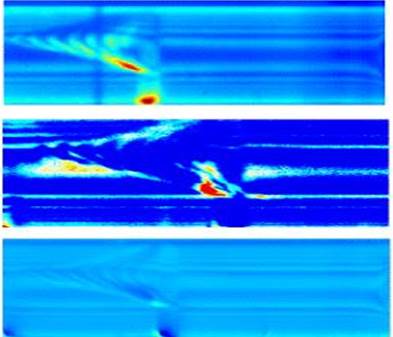
A quantitative version of “monochromatic topography” known as ‘Rocking Curve Imaging’ (RCI) was implemented, by using synchrotron light and taking advantage of the dramatic improvement of the 2D-detectors and computerised processing. The rough data is constituted by a number (~ 200) of images recorded along the diffraction (“rocking”) curve. If the quality of the crystal is such that a one-to-one relation between a pixel of the detector and a voxel within the crystal can be established (this approximation is very well fulfilled if the local mosaic spread of the voxel is < 1 mradian), RCI provides, from the each rocking curve recorded on each of the pixels of the detector, not only the “voxel” integrated intensity (the only data provided by the previous techniques), but also its “mosaic spread” (FWHM) and peak position. We will show, based on many examples, that this new data, never recorded before, open the field to a highly enhanced characterization of the crystal and deposited layers. These examples include the characterization of screw dislocations and twins occurring during silicon growth, various growth features in Al203, GaN and CdTe (where the diffraction displays the Borrmann anomalous absorption, which leads to new type of images), and the characterisation of the defects within deposited layers, or their effect on the substrate.
The figure shows a “section topograph” of a nearly perfect crystal, which exhibits Kato’s interference fringes predicted by dynamical theory. The sensitivity of the RCI technique is such that it can reveal very small modifications (that were unexpected for us) of the local FWHM and peak position (of the order of the μradian) at the level of these fringes.
1 A reference book for dynamical theory, Borrmann effect and diffraction topographic techniques (excluding RCI) is: André Authier, Dynamical Theory of X-Ray Diffraction, Oxford University Press (2001)