Séminaire - 16/03/2023 - Edoardo Zatterin - Spatially resolved crystallography: scanning X-ray diffraction microscopy at the synchrotron
Ce séminaire sera également accessible en visio (détails à la fin de ce mail).
Invitation : Thomas Cornelius (Département PHANO, Equipe MNO).
Diffusion : IM2NP, CINaM, Irphe, LP3 (via N. Sannier), Madirel (via P. Boulet), PIIM (via T. Angot), CPT (T. Martin), Fédération de Chimie (via S. Viel), CP2M
SEMINAIRE Jeudi 16 Mars 2023 à 14h00
Salle des séminaires de l'Im2np, campus de Saint-Jérôme, 1er étage Bâtiment Poincaré
et sur zoom: https://univ-amu-fr.zoom.us/j/82408267498?pwd=NUtRZnY1MTMzbzh3TzNzazlRcGJUUT09
Edoardo Zatterin
ID01, ESRF - The European Synchrotron, 71 Avenue des Martyrs, CS40220, 38043 Grenoble Cedex 9, France
Spatially resolved crystallography: scanning X-ray diffraction microscopy at the synchrotron
The study of correlations between atomic structure and macroscopic physical properties is essential to both fundamental and applied research in solid state physics. Conventional X-ray Bragg diffraction excels in this task, albeit being limited in that only the structure
corresponding to the average of the irradiated volume can be extracted. This limitation is partly overcome by scanning probe techniques, capable of determining structural parameters with spatial resolution; however, these are normally either destructive (e.g., TEM) or
surface-limited (e.g., AFM).
Thanks to the high photon flux available at synchrotron sources, X-rays can be focused by reflective or diffractive optics in sub-100nm2 spot sizes, effectively creating “X-ray nanoprobes”. By raster scanning such nanoprobes on a crystal, quantities typically discerned
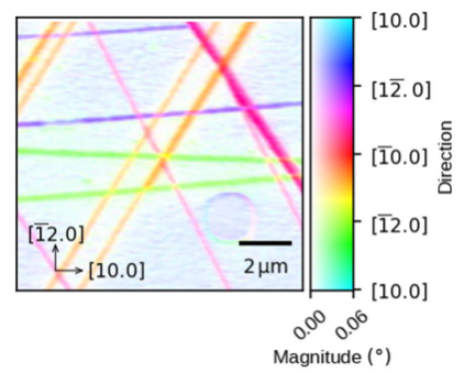
from X-ray diffraction experiments can be measured locally. Namely, one can obtain spatial maps of lattice parameters and rotations, non-destructively and in buried layers. The procedure is referred to as scanning X-ray diffraction microscopy (SXDM).
Despite being in its infancy, SXDM experiments can be performed at different synchrotrons worldwide [1,2,3,4], and the community exploiting it is growing. For example, SXDM has been used to map the crystallography and dynamics of magnetic [5], ferroelectric [6], and ferroelastic domains [7], as well as to measure the lattice displacement induced by GHz surface acoustic waves [8]. Most interestingly, in its most comprehensive form SXDM is capable of measuring the full strain tensor of the mapped sample area [9].
In this talk, I will present the working principle of SXDM and discuss its applications. Using many examples, I will show how a typical experiment is performed and what local crystallographic parameters are extracted from it.
References :[1] Leake, S. J., et al. (2019) Journal of Synchrotron Radiation, 26(2), 571–584. (ESRF)
[2] Winarski, R. P., et al. (2012) Journal of Synchrotron Radiation, 19(6), 1056–1060. (APS)
[3] Johansson, U., et al. (2021) Journal of Synchrotron Radiation, 28(May), 1935–1947. (MAX IV)
[4] Nazaretski, E., et al. (2017) Journal of Synchrotron Radiation, 24(6), 1113–1119. (NSLS-II)
[5] Evans, P. G., et al. (2020) Science Advances, 6(40), 1–8.
[6] Hadjimichael, M., et al. (2018) Physical Review Letters, 120(3), 037602.
[7] Marçal, L. A. B., (2020) ACS Nano, 14(11), 15973–15982.
[8] Hanke, M., (2023) Physical Review Applied, 19(2), 024038.
[9] Corley-Wiciak, C., et al. (2023) ACS Applied Materials & Interfaces 15 (2), 3119-3130